


How Ultrasound Imaging Changes the Way TMJ Is Diagnosed
Jaw pain has a way of affecting much more than your ability to chew. It can make conversations uncomfortable, turn meals into a challenge, trigger headaches, and even interfere with sleep. Because the temporomandibular joint (TMJ) is one of the most frequently used joints in the body, even small problems can have a noticeable impact on daily life. The challenge is that TMJ disorders don’t always have a single, obvious cause, making an accurate diagnosis one of the most important steps toward meaningful relief.
Advances in diagnostics, particularly ultrasound imaging, are changing the way healthcare providers evaluate TMJ disorders. Rather than relying solely on a physical examination or a patient’s description of symptoms, ultrasound provides a real-time view of the muscles, tendons, and joint structures involved in jaw movement. This technology allows providers to gather valuable information that supports more personalized treatment decisions and, when appropriate, guides targeted procedures with greater precision.
If you’ve been searching for answers to persistent jaw pain, continue reading to learn how ultrasound imaging is transforming TMJ diagnosis and why this technology is becoming an important part of modern patient care.
What Is Ultrasound Imaging?
Before exploring its role in TMJ care, it’s helpful to understand how ultrasound imaging works. Ultrasound imaging is a non-invasive diagnostic technology that uses high-frequency sound waves to create real-time images of structures inside the body.
While many people associate ultrasound with pregnancy, the same technology is widely used to evaluate muscles, tendons, ligaments, joints, and other soft tissues without exposing patients to radiation.
For TMJ evaluation, ultrasound allows providers to visualize the jaw joint and surrounding tissues as they move. This dynamic view can provide valuable information about how the joint functions, helping identify areas of inflammation, muscle involvement, or structural abnormalities that may contribute to discomfort.
Ultrasound imaging offers several advantages, including:
- Real-time visualization of the jaw joint during movement
- A non-invasive examination without radiation exposure
- Detailed evaluation of muscles, tendons, and soft tissues
- Support for more accurate TMJ imaging diagnosis
- Guidance for certain in-office procedures when appropriate
Because ultrasound produces immediate images, providers can often correlate what they see with the patient’s symptoms during the same appointment, making it a valuable tool in TMJ diagnostic technology.

How Ultrasound Imaging Changes the Way TMJ Is Diagnosed
Diagnosing TMJ disorders isn’t always straightforward. Jaw pain may originate from the joint itself, surrounding muscles, tendons, or a combination of factors. Ultrasound imaging gives providers another layer of information, allowing them to evaluate these structures with greater confidence before developing a treatment plan.
1. See the Joint in Real Time
One of ultrasound’s greatest advantages is its ability to show movement as it happens. Unlike static images, ultrasound allows providers to observe the temporomandibular joint while a patient opens and closes the mouth.
Watching the joint move in real time can reveal patterns that may not be apparent during a routine physical examination alone. This dynamic evaluation helps providers assess how the joint and surrounding tissues function together rather than relying solely on symptoms or external observations.
For patients experiencing clicking, popping, limited jaw movement, or discomfort while chewing, this real-time perspective can provide valuable insight during a TMJ evaluation in NJ.
2. A More Complete Diagnosis
TMJ disorders can present with many different symptoms. Some patients experience jaw pain, while others primarily notice headaches, facial tension, ear discomfort, or difficulty opening the mouth fully. Because these symptoms overlap with several other conditions, accurate diagnosis is essential.
Ultrasound imaging contributes to a more comprehensive evaluation by allowing providers to examine soft tissue structures that cannot be assessed through physical examination alone. Combined with a patient’s medical history and clinical assessment, ultrasound can assist in evaluating:
- Muscle inflammation
- Tendon abnormalities
- Joint irritation
- Soft tissue changes
- Areas of tenderness corresponding with symptoms
This additional information strengthens the overall TMJ imaging diagnosis, helping providers better understand the source of discomfort before recommending treatment.
3. Better Information Leads to Better Planning
Every TMJ disorder is different. Some patients have symptoms primarily related to muscle tension, while others have inflammation within the joint or mechanical dysfunction affecting jaw movement. Because the underlying cause can vary, treatment should never be based on assumptions alone.
Ultrasound imaging supports individualized care by helping providers gather more precise diagnostic information before creating a treatment plan. Instead of treating symptoms alone, providers can better determine which therapies are most appropriate based on what they observe during the evaluation.
For patients seeking answers for persistent imaging for jaw pain, this technology helps create a clearer understanding of what’s happening beneath the surface.
4. Greater Precision During Injections
Ultrasound doesn’t only assist with diagnosis. It also plays an important role when image-guided procedures are recommended. During an ultrasound-guided TMJ injection, real-time imaging allows the provider to visualize the needle as it approaches the targeted structure.
Rather than relying solely on surface landmarks, ultrasound provides continuous guidance throughout the procedure. This approach may improve guided injection accuracy by helping providers place medication in the intended location while avoiding nearby structures.
For patients who are candidates for injection therapy, ultrasound guidance represents an important advancement in both precision and confidence during treatment.
5. It Helps Connect Symptoms With What Providers See
Many patients describe TMJ symptoms that seem inconsistent from day to day. One morning, the jaw feels stiff. Another day, chewing becomes uncomfortable. Headaches may appear without warning, or clicking may occur only during certain movements.
Ultrasound imaging allows providers to compare what patients are experiencing with what is happening inside the joint during those same movements. This real-time connection between symptoms and imaging often provides valuable context during the diagnostic process, helping providers develop treatment recommendations that are based on objective findings rather than symptoms alone.
6. A Comfortable, Non-Invasive Experience
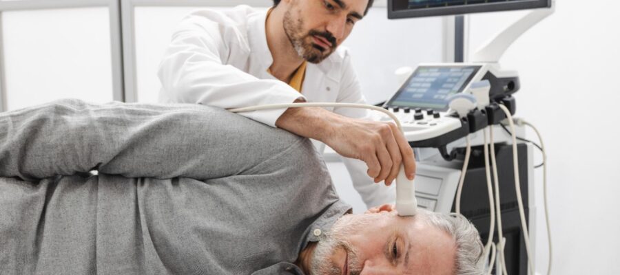
For many patients, one of the most appealing aspects of ultrasound imaging is its simplicity. The examination is performed externally using a handheld device that glides gently over the skin near the jaw. There are no incisions, needles, or radiation involved during the imaging itself, and the procedure is generally well tolerated.
Because images appear immediately, providers can evaluate jaw movement, discuss findings, and answer questions during the same visit. For patients exploring diagnostic imaging for jaw pain, this combination of comfort and immediate feedback makes ultrasound an increasingly valuable part of modern TMJ care.
7. Monitoring Progress Over Time
Diagnosing TMJ disorders is only the beginning of the treatment journey. Because symptoms can change as treatment progresses, providers often benefit from tools that allow them to evaluate the joint over time. Ultrasound imaging makes this possible by providing repeatable, real-time views of the jaw without exposing patients to radiation.
Follow-up imaging may help providers:
- Observe changes in inflammation
- Assess how the joint responds to treatment
- Compare findings from previous visits
- Support adjustments to the treatment plan when needed
Rather than relying only on how a patient feels, providers can combine symptom improvement with imaging findings to build a more complete picture of progress. This ongoing evaluation helps create a treatment plan that’s responsive to each patient’s needs.

Better Diagnosis Leads to Better Care
Persistent jaw pain deserves more than guesswork. Modern diagnostics, including ultrasound imaging, allow healthcare providers to evaluate the temporomandibular joint in real time, providing valuable insight into how the joint and surrounding soft tissues function.
By supporting a more accurate TMJ imaging diagnosis and improving precision during procedures such as an ultrasound-guided TMJ injection, this technology helps create treatment plans that are tailored to each patient’s unique condition.
At the Headache & TMJ Center of New Jersey in West Orange, NJ, advanced diagnostic technology is an important part of delivering personalized TMJ care. If jaw pain, headaches, or facial discomfort have been affecting your daily life, a comprehensive evaluation that includes modern imaging may be the first step toward understanding the cause and identifying the treatment approach that’s right for you.
Call us today to schedule your appointment!
